Love Bridge workflow
Was just going through tons of pictures tonight and this is a case I finished a couple weeks ago. I don't do many bridges, but you have have to love the workflow when you do them.
- Prep Abutment teeth
- Image preps and root
- Mill Teliocad Temp and extract root while milling
- Reline pontic to form ovate pontic and cement
- After 10 week healing time restore...
Works beautifully... Also, if you have the 4 motor mill, use EF milling. Takes a long time, but the mills are phenomenal.
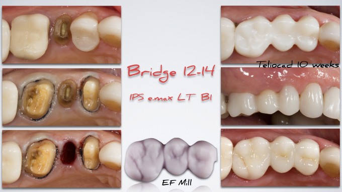
btw.. just the bottom right is immediate final picture... I don't have final pictures yet. The top right and the middle right are both the Teliocad Provisional at 10 weeks. I will take a healing shot at recall.
Mike, Do you rescan for the final? If not how do you account for the tissue change? If you mill the Emax bridge from the original scan do you just adjust the pontic?
On 7/18/2017 at 9:09 pm, Dhaval Patel said...Mike, Do you rescan for the final? If not how do you account for the tissue change? If you mill the Emax bridge from the original scan do you just adjust the pontic?
Hi Dhaval-
Generally I do. You could technically just go back to the old images, use the cut tool and then scan the new tissue/preps, but I find it just as easy to rescan. I scanned the Temp Bridge in a Biocopy folder (just in case), removed the temp and refined the margins of the prep (drop a little around newly formed ovate pontic) and rescanned that as well. Just took about 10 minutes for all of it.
That would be nice...
However, you could Biocopy I guess. I wanted to redesign anyway for better anatomy with the EF Mill
Very nice workflow. The margins of the temps are so much better milled than when we used to make these chairside. Tissue is so nice when you remove for rescan and at delivery. Great case to show off this technique.