Imaging with the CEREC Omnicam and Bluecam

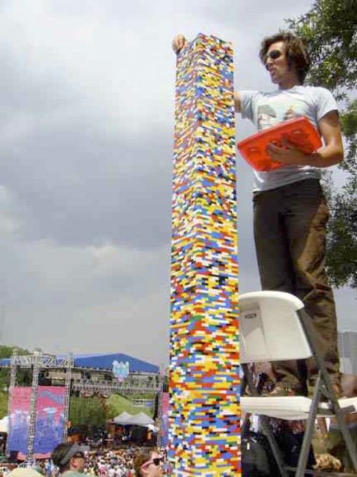
Admit it, as a kid you took a stack of Legos and stacked them as high as you could - only to see the tower as it got higher lean over and topple.
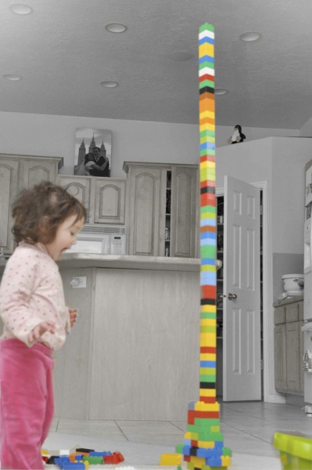
With age comes wisdom and you realize that if you wanted to stack that Lego tower as high as possible, you simply could not just stack the pieces as individual pieces. You had to crisscross them. You had to reinforce the Lego bricks to give the tower more stability. The more reinforcement, the more stable the tower.
This might be all fine and dandy but you ask, what does this have to do with imaging with the CEREC? Frankly lots. If you think about how most people image, they image in a linear fashion. They image the occlusal, the buccal and then then lingual and the model is created.
As the Omnicam takes images, those images are stacked on top of each other, not unlike a Lego brick and eventually the model is created. However, if all you do is take images in a straight line, as your model gets bigger, the model becomes more unstable.
So how should we image? Roll the camera buccally and lingually to not only capture and fill in the data, but also reinforce the model. With additional images comes additional overlapping of the images which leads to additional structural stability of the model.
All of this is probably not critical when doing a small quadrant. It becomes extremely relevant when taking a full arch scan for the virtual articulation feature or for taking scans for night guards or orthodontics.
If you want to test this theory, image a full upper arch on a model and a full lower arch. Take a buccal bite and see what happens on the contralateral side to the buccal bite if you just take linear scans. Now do the same thing and take rolling scans and you will find your articulation much more accurate.
To see a great video on the rolling scan technique, check here: http://www.cerecdoctors.com/digital-learning/view/id/945/category/177